Imaging
Differences Between Dental X-rays and Tomography
Explains how panoramic X-rays, small dental films, and dental tomography can provide different information.
Prepared by
Dt. Seçil Sönmez
Clinical review
Dt. Seçil Sönmez, Dentist
Updated
May 13, 2026
Read time
5 min
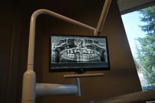
Dental X-ray is not one single type of image. Small intraoral films can show a specific tooth, root, filling, or decay relationship, while panoramic X-rays help review the jaw and multiple areas in a wider frame.
Dental tomography can provide three-dimensional information. Implant planning, impacted teeth, bone structure, sinus relationship, or complex surgical assessment may require more detail. Tomography is not necessary in every case; the need depends on examination findings.
The goal of imaging is not to take the greatest number of images, but to answer the right clinical question. Decay, root canal treatment, implants, oral surgery, and orthodontics may each require different imaging choices.
If the patient already has older images, bringing them to the appointment can help reduce unnecessary repetition. The most useful image is decided after symptoms, examination, and previous records are reviewed together.
General information
This article is for general information and does not replace a personal diagnosis or treatment plan. Dental concerns should be evaluated by a dentist.