التصوير
الفروق بين أشعة الأسنان والتصوير المقطعي
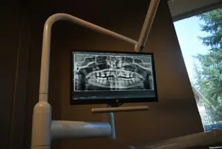
يوضح كيف تقدم الأشعة البانورامية وصور الأسنان الصغيرة والتصوير المقطعي معلومات مختلفة.
أعده
Dt. Seçil Sönmez
مراجعة سريرية
Dt. Seçil Sönmez, طبيب أسنان
آخر تحديث
13 مايو 2026
مدة القراءة
5 دقائق
Dental X-ray is not one single type of image. Small intraoral films can show a specific tooth, root, filling, or decay relationship, while panoramic X-rays help review the jaw and multiple areas in a wider frame.
Dental tomography can provide three-dimensional information. Implant planning, impacted teeth, bone structure, sinus relationship, or complex surgical assessment may require more detail. Tomography is not necessary in every case; the need depends on examination findings.
The goal of imaging is not to take the greatest number of images, but to answer the right clinical question. Decay, root canal treatment, implants, oral surgery, and orthodontics may each require different imaging choices.
If the patient already has older images, bringing them to the appointment can help reduce unnecessary repetition. The most useful image is decided after symptoms, examination, and previous records are reviewed together.
معلومات عامة
هذه المقالة مخصصة للمعلومات العامة ولا تغني عن التشخيص الشخصي أو خطة العلاج. ينبغي تقييم مشكلات الأسنان لدى طبيب أسنان.