Diagnosis and Planning
When Are Dental X-Rays and Panoramic Imaging Requested?
Explains why dental X-rays, panoramic imaging, and tomography may be considered for decay, impacted teeth, implant planning, and oral surgery.
Erstellt von
Dt. Seçil Sönmez
Klinische Prüfung
Dt. Seçil Sönmez, Zahnarzt
Aktualisiert
13. Mai 2026
Lesezeit
5 min
Imaging is not needed at every dental examination, but X-rays or panoramic imaging may be requested when information not visible to the eye is needed. Decay depth, root structure, impacted tooth position, bone support, and the condition of previous treatments are examples.
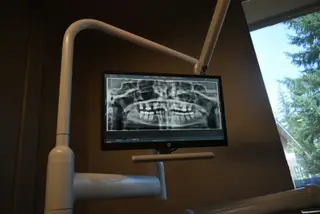
Panoramic imaging shows the mouth and jaws in a wider frame. It can help review impacted wisdom teeth, multiple missing teeth, oral surgery assessment, implant planning, or broader gum-related concerns.
Tomography is not routine for every patient. It may be considered when more detailed three-dimensional information is needed, such as bone volume around an implant area, proximity to the nerve canal, or surgical planning. The need depends on examination findings.
The purpose of imaging is not to do more treatment. It is to plan more accurately. When the complaint, clinical findings, and previous records are reviewed together, unnecessary repetition can be avoided and needed information becomes clearer.
Allgemeine Information
Dieser Artikel dient der allgemeinen Information und ersetzt keine persönliche Diagnose oder Behandlungsplanung. Zahnmedizinische Beschwerden sollten zahnärztlich beurteilt werden.